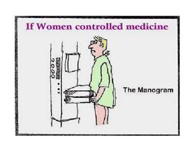
 Do you regularly have breast cancer screening – are the mammograms you have safe? Have you been told, before the procedure, about the side effects of mammograms? Have you been told how effective at reducing breast cancer mammograms are? The fact is, mammograms do more harm than good!
Not what conventional medicine says.
But that’s not what conventional medicine tells you. Mammograms are said to reduce the risk of dying from breast cancer by an incredible 20%! But how has that figure been derived?
Let’s work it out!
This 20% actually amounts to just 1 woman in 1,000 who gets regular mammograms over a lifetime. But for every 1,000 women who don’t have mammograms, 5 of them will die of breast cancer. This means that of those who do get the procedure, 4 will die anyway. So how do they get to 20%? Well, the difference between the two groups. The 5 that will die without mammograms versus the 4 that will die with mammograms, is 20%! Doh!!
Here’s a video to make is clearer:
Manipulated figures
This is how figures are manipulated to be in favour of the treatment or drug. In this case, it amounts to 1 woman in 1,000 being saved by regular mammograms. But wait a minute, what about the side effects.
What about the false positives?
Going back to our 1,000 women, half of these will receive a false positive. In other words, although they don’t have cancer, they will go through the torment and horror of thinking they do have it.
…and biopsies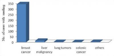
64 of these terrified women will have biopsies which in themselves carry high risks of adverse effects. 10 of these women will go on to have cancer treatment for a disease they haven’t got!
The majority of medical professionals will tell you there is little risk of ‘seeding’ of cancer cells from a breast biopsy. But have a look at the bar chart from research done in 2014. Also another research paper concludes that breast biopsies can cause metastases.
…and unnecessary treatment?
This will include disfiguring surgery and toxic chemo and/or radiation. These three methods of diagnosing and treating cancer are all extremely risky and carcinogenic in their own right. Dying from the treatment of a disease you didn’t have in the first place, is a crime to say the least. If you don’t die, these treatments can make you sick and cause metastases.
10 healthy women out of 1,000 are treated erroneously!
This means 10 women out of 1,000 have cancer treatment when they have nothing wrong with them. Apart from that, they may even contract cancer from the toxic and carcinogenic treatment they have erroneously received and could die from these unwarranted procedures.
Dr Karsten Juhl Jorgensen
Dr. Karsten Juhl Jorgensen, deputy director of the Nordic Cochrane Center:
“Some types of screening are a good idea … But breast cancer has a biology that doesn’t lend itself to screening.
Healthy women get a breast cancer diagnosis, and this has serious psychological consequences and well-known physical harms from unnecessary treatment. We’re really doing more harm than good.”
$4 billion on false-positive mammograms
A study published in “Health Affairs” in 2015 stated that a massive $4 billion was spent each year on false-positive mammograms. These would lead to breast cancer overdiagnosis amongst women from the ages of 40 through to 59. What a waste of money but more importantly, how does this affect those women who are misdiagnosed.
 Fran Visco
Fran Visco, President of the National Breast Cancer Coalition (NBCC) told Newsweek “getting unnecessary treatment for harmless tumours can jeopardize health. Something that women are rarely informed about.” She also cited that breast cancer activist Carolina Hinestrosa, a vice president at the coalition, died at age 50 from soft-tissue sarcoma, a tumor caused by radiation used to treat an early breast cancer. Visco continued “Women should understand these risks. Instead, women often hear only about mammograms’ benefits. Women have been inundated with the early-detection message for decades.”
Mammograms are X-rays
There is no real way of telling whether the regular mammogram that women are receiving is actually causing cancer. We all know that X-ray radiation has risks and women are having regular doses of it.
Women are so trusting!
Women need to stop trusting their doctors or oncologists until they have researched their own disease first. Why are we putting our bodies in the hands of people that don’t even know us. Medical professionals are constantly being advised and cajoled by the pharmaceuticals, whose only interest is profits. Doctors are forever short of time, with only a few minutes available to assess our condition before moving onto the next number.
 Check your diet for Calcium
Mammography invariably find small anomolies such as scattered calcifications, which can take years to form.
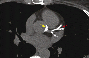
Protect yourself by not taking calcium (Ca) supplements. We get enough Ca from our diet. Too much Ca or too little magnesium (Mg) will give you the same symptoms. These two vital minerals have to be in equilibrium to work optimally. With our modern diet, Ca is in abundance and Mg is scarce, hence many health problems, such as this image showing the arteries of the heart being blocked by calcifications.
Don’t panic!
If you do have cancer, don’t panic! it has probably been forming for years. You have time to do your own research and be educated enough to make an informed decision as to what treatment you want. Always look at natural alternatives; they will have no side effects but are often more affective than toxic treatments. Chemo and radiation therapy can have affects on your body far into the future. If in doubt, it is your prerogative to get a second opinion on your diagnosis.
Harvard Medical School
According to the Harvard Medical School: “The radiation you get from x-ray, CT, and nuclear imaging is ionizing radiation — high-energy wavelengths or particles that penetrate tissue to reveal the body’s internal organs and structures. Ionizing radiation can damage DNA, and although your cells repair most of the damage, they sometimes do the job imperfectly, leaving small areas of “misrepair.” The result is DNA mutations that may contribute to cancer years down the road.”
Benign Calcifications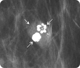
In this image a fibroadenoma (arrows) is present with dense, coarse calcifications (double arrows). These types of calcifications are distinctive and when seen, whether alone or in a mass, a biopsy is not needed.
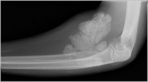
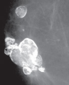 The next image shows the result of a car accident which has produced large calcifications due to trauma. No treatment was necessary.
Ductal Carcinoma in Situ (DCIS)
An estimated 33% of new diagnoses which are obtained through a mammogram, are classified as ductal carcinoma in situ (DCIS). This applies to abnormal growth of cells in the milk ducts. These lesions are small at around 1cm in diameter. They can appear as a malignant growth under the microscope but they have not invaded any surrounding tissue.
DCIS diagnosis at epidemic proportions
As a consequence DCIS is appraised as a stage zero breast cancer. Some would argue that it needs to be considered as a non-cancerous condition. If it weren’t for the use of X-ray diagnostics, this would not even be discovered. In fact, in the 1980s, when more emphasis was put on breast cancer awareness, the diagnosis of DCIS began to increase to our present day epidemic proportions.
 According to the ACS
According to the American Cancer Society (ACS), this year (2017) will see an estimated quarter million new breast cancer cases diagnosed in the U.S. It is also estimated that around 41,000 women will die from breast cancer.
When in doubt, cut it out!
An additional estimated 63,000 women will be diagnosed with DCIS. Most of these patients will undergo the exact same treatment as those with invasive breast cancer. The reason for this is because doctors typically recommend treating DCIS in order to prevent it from becoming invasive. In other words, a mentality of “when in doubt, cut it out” tends to predominate. However, when you consider the risks of serious harm — including death — from the treatment itself, is this really the wisest strategy?
Newsweek:
“Other experts note that DCIS carries such low risk that it should be considered merely a risk factor for cancer. Researchers are conducting studies to measure whether it’s safe to scale back treatment of DCIS … In the meantime, [patients] and their doctors must make difficult choices without knowing for sure whether it’s the right thing to do.”
Other considerations with diagnosis
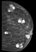
As we get older, most women get calcifications within the breast tissue. This can be seen by a mammogram X-ray. These calcifications are mostly harmless.
2 main types of breast calcifications:
- Macrocalcifications – randomly dispersed large white dots. These are found in around half of women over the age of 50, and about 10% of women under age 50. They are considered noncancerous and need no further treatment.
- Microcalcifications – these look like white specks on a
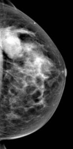
Benign fibroadenoma mammogram that often appear is clusters. This is not likely to be cancer, but your doctor may want another mammograph or a biopsy to double check. Some doctors can be eager to do something, just in case.
But this is your body and it is your prerogative to make an informed decision. Biopsies, especially with the breast tissue, have their own risks. See the bar chart above.
If it were me, I would definitely be wary of having any treatment for a ‘just in case’ scenario. But that’s just my personal opinion. Don’t forget, benign calcifications usually get worse as we age, but you can do something about that!
Vitamin K2
We shouldn’t just accept these Ca deposits. They are a warning that we have excess Ca in our bodies and their is a solution. There is a little known molecule known as vitamin K2. This vitamin is necessary to activate MGP (matrix gla protein), the Gla is short for glutamic acid. If you are deficient in K2, MGP will not do its job of ushering excess Ca out of the soft tissue and into the bones. Osteocalcin, which resides in the bone, then keeps Ca there where it belongs.
A great combination
MGP and osteocalcin are both vitamin K2 dependent and neither can function without it. Mg, MGP, K2 and osteocalcin are a great combination to reduce calcification of your tissues. 99% of Ca should reside in the bones. The sunshine vitamin D3 will also help this process, but be careful not to overdose on D3 supplements. Sunshine is best, if you can get it!
A bonus!
These nutrients help with calcifications all over the body, including the heart, arteries, kidney stones, periferal artery disease, heel spurs etc. Reduce your calcification burden with a good quality supplement of Mg and K2. Get your Ca from diet only. Don’t take Ca supplements, we are already overloaded with fortified Ca foods. Get as much natural sunshine as you can. Keep your health protected for now and in the future.
 Incidently
Vitamin D3 (which is actually a hormone), needs Mg to activate it as it is stored in the body in its inactive form. Another reason to keep your Mg levels up. Without it, your D3 levels can appear low when in fact is is just inactivated.